About Fibralign
Company Overview

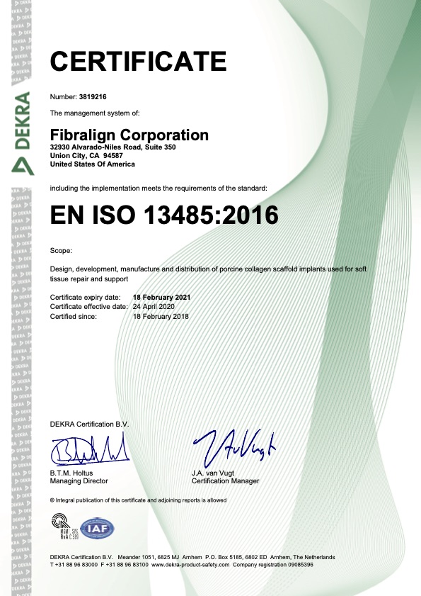
Fibralign is an award-winning, commercial-stage, Stanford-spinout that designs, develops and produces novel therapeutic medical devices to address major unmet medical needs. Based in the San Francisco Bay Area, the company’s team has established GMP commercial production (ISO 13485:2016 certified), quality system and development labs co-located at its main facility (Union City, CA).
Fibralign is supported in its mission by meaningful collaborations with leading research universities, renowned medical leaders and strategic partnerships. These relationships have proven invaluable in advancing research and development, resulting in a compelling pipeline of novel products that are based on Fibralign’s proprietary Nanoweave® scaffolding platform.
Privately held, Fibralign has been able to accomplish this progression with the support of investors and grant funding from the Department of Defense, National Institutes of Health and the National Science Foundation.
Fibralign won the prestigious Medtech Innovator Award, taking first out of over 300 startups that participated in this annual competition. The team has maintained a strong relationship with its Stanford roots, working closely with several research groups in preclinical and clinical studies and as an active member of the Stanford StartX community. Fibralign is also an alumnus of JLABS , having been selected to join in the initial cohort of startup members for its South San Francisco location (JLABS@SSF).
Nanoweave® Technology

Fibralign’s patented Nanoweave® technology provides the means to induce self-assembly of molecular type I collagen to replicate native tissue scaffold outside of the body. This fundamental breakthrough enables medical grade collagen to be precisely printed with nanoscale features that mimic human tissue – to such a degree that the body’s tissue will interact with it naturally and not reject or challenge it as a foreign object. Multiple studies (Stanford, UCSF, others) have shown that fibroblasts, myoblasts, and mesenchymal stem cells proliferate, migrate and align on these fabricated matrices. Preclinical studies have also demonstrated that Nanoweave matrices induce remodeling phenotype, supporting the body’s regenerative repair and maintenance process.
There is increasing evidence, as shown in peer review articles, that the efficacy of tissue regeneration depends on creating a suitable micro-environment that can support cell function and decrease pathological healing. Nanoweave successfully mimics the targeted tissue, both in bio-mechanical properties and biochemical composition, and provides the appropriate cell-to-matrix interactions, adhesion, proliferation, maturation, etc.
Fibralign’s core technology provides a rich platform for scientific research and commercial development in the fields of tissue engineering and regenerative medicine. BioBridge represents just the first product commercialized using Nanoweave technology. The company’s development efforts have provided deep experience and an extensive “tool box” for application development, which has been validated in preclinical models through university research collaborations for a wide range of potential novel therapies. This has resulted in the development of a compelling pipeline of addressing other significant medical conditions that where lymphatic impairment contributes or causes chronic conditions, including neurological disorders, heart disease and glaucoma.


Publications
Over 40 peer-reviewed papers have been published regarding BioBridge preclinical and clinical findings, with over 25 additional publications related to Fibralign’s BioBridge® and Nanoweave® technology.
A subset of selected papers are listed below:
- Bae A, Loening A, Carrion K, Posternak V, He H, Leong S, Nguyen, D. Lymphatic regeneration visualized on MRI after placement of aligned nanofibrillar collagen scaffolds for treatment of lymphedema. Ann Plast Surg. 2025 Jun 13.
- Lazar SV, Song S, Eggert GR, Cheng MH, Nguyen DH. Single-Malt Whisky Versus Cocktail? Approaches to Surgical Lymphedema Management. J Surg Oncol. 2025 Jan 05
- Dimitrios Dionyssiou, Antonios Tsimponis, Eleni Georgiadou, Konstantina Mamaligka, Efterpi Demiri. Long-term results of lymphedema treatment with Combined lymph node transfer and collagen scaffolds: An Observational Study. JPRAS. Open 43 (2025) 328-339.
- Barrantes S, de la Torre S, Visconti G, Blasi M, López-Ojeda A. Treatment of Genital Lymphedema With Lymphatic System Pedicled SCIP Transfer in Combination With Nanofibrillar Collagen Scaffolds. Plast Reconstr Surg Glob Open. 2024 Nov;12(11):e6335.
- Mohan AT and Nguyen DH. BioBridge in Lymphatic Surgery. Chapter in book by Giuseppe Visconti et al. (Eds): Supermicrosurgical Lymphaticovenular Anastomosis. Springer Nature. 2024.
- Drobot D, Leitner Shemy O, Zeltzer AA. Biomaterials in the clinical treatment of lymphedema – a systematic review. J Vasc Surg Venous Lymphat Disord. 2024 Jan;12(1):101676.
- Szymanski K, Chun Fat S, Brazio PS. Surgical Treatment of Breast Lymphedema: A Distinct Pathology With Unique Challenges. Ann Plast Surg. 2024 May 01;92(5S Suppl 3):S315-S319.
- Chaker SC, James AJ, King D, Karagoz H. Lymphedema: Current Strategies for Diagnostics and Management. Ann Plast Surg. 2024 Dec 01;93(6S Suppl 3):S167-S171.
- Salibian AA, Yu N, Patel KM. Staging Approaches to Lymphatic Surgery: Techniques and Considerations. J Surg Oncol. 2024 Nov 18
- Cè M, Menozzi A, Soresina M, Giardini D, Martinenghi C, Cellina M. Lymphedema Surgical Treatment Using BioBridge: A Preliminary Experience. Appl. Sci. 2023, 13, 11571
- Campos JL, Pons G, Al-Sakkaf AM, Luse IL, Pires L, Vela FJ, Ramos E, Crisóstomo V, Sánchez-Margallo FM, Abellán E, Masiá J. Lymphatic Regeneration after Popliteal Lymph Node Excision and Implantation of Aligned Nanofibrillar Collagen Scaffolds: An Experimental Rabbits Model. J. Funct. Biomater. 2024, 15, 235.
- Witt M., Ring A., Stricker I., Fruth E. The Fate of Implanted BioBridge Collagen Matrix – Findings in Histology, Scanning, and Transmission Electron Microscopy. Lymphology 56 (2023) 121-124
- Nguyen D, Dionyssiou D, Zaitseva TS, Zhou AT, Sue G, Deptula P, Moroz MA, Tabada P, Rockson SG, Paukshto MV, Cheng MH, Huang NF. Development of a rat model of lymphedema and the implantation of a collagen-based medical device for therapeutic intervention. Front Cardiovasc Med. 2023;10:1214116.
- Dimitrios Dionyssiou, Dung Nguyen, Anastasios Topalis, Peter Deptula, Michael Paukshto, Tatiana Zaitseva, Efterpi Demiri, Aggeliki Cheva, Stanley Rockson. Treatment of Rat Lymphedema by Propeller Lymphatic Tissue Flap Combined with Nanofibrillar Collagen Scaffolds. J Reconstr Microsurg. 2024 Feb;40(2):145-155.
- Bernas, M., Al-Ghadban, S., Thiadens, S.R.J., Ashforth K, Lin WC, Safa B, Buntic R, Paukshto M, Rovnaya A, McNeely ML. Etiology and treatment of cancer-related secondary lymphedema. Clin Exp Metastasis Springer Nature, 2023 Sep 30. https://doi.org/10.1007/s10585-023-10232-8
- Bianchi LMG, Irmici G, Cè M, D’Ascoli E, Della Pepa G, Di Vita F, Casati O, Soresina M, Menozzi A, Khenkina N, Cellina M. Diagnosis and Treatment of Post-Prostatectomy Lymphedema: What’s New? Curr Oncol. 2023 Apr 25;30(5):4512-4526.
- Inchauste SM, Nguyen DH, and Ming-Huei Cheng. Animal Study and Cadaver Dissection of Lymphedema. In book: Principle of Lymphatic System. Elsevier, 2022.
- Chin‐Yu Yang, Ines E. Tinhofer, Dung Nguyen, Ming‐Huei Cheng. Enhancing lymphangiogenesis and lymphatic drainage to vascularized lymph nodes with nanofibrillar collagen scaffolds. J Surg Oncol. 2022;1‐
- Deptula P, Zhou A, Posternak V, He H, Nguyen D. Multimodality Approach to Lymphedema Surgery Achieves and Maintains Normal Limb Volumes: A Treatment Algorithm to Optimize Outcomes. J Clin Med. 2022 Jan 25;11(3):598.
- Nguyen DH, Zhou A, Posternak V, Rochlin DH. Nanofibrillar Collagen Scaffold Enhances Edema Reduction and Formation of New Lymphatic Collectors after Lymphedema Surgery. Plast Reconstr Surg. 2021 Dec 1;148(6):1382-1393.
- Nguyen D, Zaitseva TS, Zhou A, Rochlin D, Sue G, Deptula P, Tabada P, Wan D, Loening A, Paukshto M, Dionyssiou D. Lymphatic regeneration after implantation of aligned nanofibrillar collagen scaffolds: Preliminary preclinical and clinical results. J Surg Oncol. 2021 Sep 21. doi: 10.1002/jso.26679. Epub ahead of print. PMID: 34549427. https://pubmed.ncbi.nlm.nih.gov/34549427/
- Dionyssiou, Dimitrios & Demiri, Efterpi. (2021). A comprehensive treatment algorithm for patients requiring simultaneous breast and lymphedema reconstruction based on lymph node transfer. Annals of Breast Surgery. 10.21037/abs-20-142.
- Zaitseva T, Yang G, Dionyssiou D, et al. Delivery of hepatocyte growth factor mRNA from nanofibrillar scaffolds in a pig model of peripheral arterial disease. Regen Med. 2020;15(6):1761-1773. doi:10.2217/rme-2020-0023 [https://pubmed.ncbi.nlm.nih.gov/32772903/]
- Hu C, Zaitseva TS, Alcazar C, et al. Delivery of Human Stromal Vascular Fraction Cells on Nanofibrillar Scaffolds for Treatment of Peripheral Arterial Disease. Front Bioeng Biotechnol. 2020;8:689. Published 2020 Jul 17. doi:10.3389/fbioe.2020.00689 [https://doi.org/10.3389/fbioe.2020.00689]
- Rochlin, D, Inchauste S, Zelones J, Nguyen DH. The role of adjunct nanofibrillar collagen scaffold implantation in the surgical management of secondary lymphedema: Review of the literature and summary of initial pilot studies. Journal of Surgical Oncology, 2020. 121(1): p. 121-128 [https://onlinelibrary.wiley.com/doi/full/10.1002/jso.25576]
- Inchauste S, Zelones J, Rochlin D, Nguyen DH. Successful treatment of lymphedema in a vasculopath and neuropathic patient. J Surg Oncol. 2020;121(1):182-186. doi:10.1002/jso.25590 [https://pubmed.ncbi.nlm.nih.gov/31228351/]
- Rockson SG. Lymphedema after Breast Cancer Treatment. N Engl J Med. 2018;379(20):1937-1944. doi:10.1056/NEJMcp1803290 [https://pubmed.ncbi.nlm.nih.gov/30428297/]
- Hadamitzky, C., Zaitseva, T. S., Bazalova-Carter, M., Paukshto, M. V., Hou, L., Strassberg, Z., et al. (2016). Aligned nanofibrillar collagen scaffolds – Guiding lymphangiogenesis for treatment of acquired lymphedema. Biomaterials 102, 259–267. doi: 10.1016/j.biomaterials.2016.05.040 [https://pubmed.ncbi.nlm.nih.gov/27348849/]
- Nakayama KH, Hong G, Lee JC, Patel J, Edwards B, Zaitseva TS, Paukshto MV, Dai H, Cooke JP, Woo YJ, Huang NF. Aligned-Braided Nanofibrillar Scaffold with Endothelial Cells Enhances Arteriogenesis. ACS Nano. 9(7):6900-8 (2015). doi: 10.1021/acsnano.5b00545. Epub 2015 Jun 17. [https://pubmed.ncbi.nlm.nih.gov/26061869/]
- Huang NF, Okogbaa JN, Lee JC, Paukshto M, Zaitseva T, Cooke JP. The modulation of endothelial cell morphology, function, and survival using anisotropic nanofibrillar collagen scaffolds. Biomaterials. 34:4038-4047. (2013) [https://pubmed.ncbi.nlm.nih.gov/23480958/]
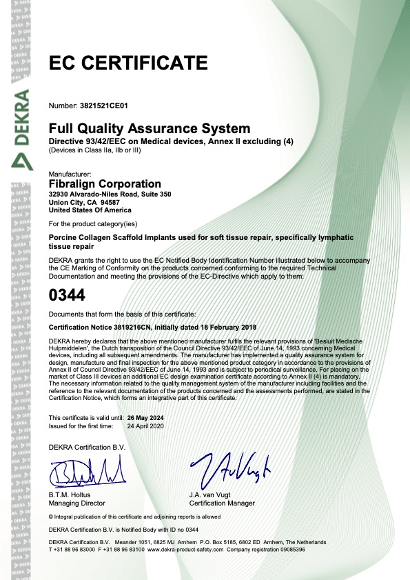
Certifications